|
published by Ecco/Harper Collins, 2006
|
|
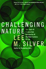
Challenging Nature
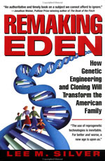
Previous books
Presentations
Publications/Writings
Biography
Princeton teaching
|
Autumn Books
Nature 431, 905-907 (21 October 2004) | doi: 10.1038/431905a
Stalking life's second secret
Lee M. Silver
BOOK REVIEWED
-
The Proteus Effect: Stem Cells and their Promise for Medicine
by Ann B. Parson
Joseph Henry Press: 2004. 304 pp. $24.95
On 28 February 1953, Francis Crick famously burst into a Cambridge pub with Jim Watson in tow and exclaimed triumphantly: "We have found the secret of life!" The rest, as the cliché goes, is history. Both scholarly and popular accounts of the personalities and events surrounding the discovery of the double helix, and subsequent advances in molecular genetics, have appeared in tens of thousands of books and articles. The DNA story is taught in school biology classes to eight-year-old children. Several DNA-based Nobel prizes have already been won, and more are no doubt waiting in the wings.
Genes and genomes, however, represent only one of two pillars of likely twenty-first-century progress in biomedicine. The second is stem-cell biology. And yet, while everyone knows of Watson and Crick, hardly anyone has heard of Leroy Stevens and other early stem-cell pioneers. Indeed, many scientists are unaware of the five decades of advances that occurred before the 1998 isolation of human embryonic stem cells. In her new book The Proteus Effect, journalist Ann Parson fills the gap with a breezy, easily accessible narrative of the people, results and ideas that have shaped the field.
Parson traces the birth of modern stem-cell research back to 1953, the same year that the double helix made its debut. Although the means for composing and reproducing life's genetic recipes had been a fundamental secret of life, it was not the only one. A second mystery was the process by which a microscopic embryo could develop into a fully functional human being or other mammal. Embryologists knew that development occurred through the controlled growth, division and specialization of individual cells, but they didn't know how developmental control was asserted. Many assumed, without evidence, that a permanent loss of biological components or capabilities was responsible for the narrowing of cellular potential. By implication, it seemed that a postembryonic mammalian cell should never be able to reverse course and return to an embryonic state. The exceptions were the germ cells (eggs and sperm), which still had the potential to retrace normal development only after combining with each other.
Goal-oriented unidirectional development was easily accepted as scientific dogma because it aligns neatly with traditional Judaeo-Christian-derived religious doctrine, which permeates Western culture. As recently as 2002, Princeton professor Robert George — a defender of strict Catholicism and member of President Bush's Council on Bioethics — claimed the mantle of science when he wrote that the direction of a human embryo's growth "is not extrinsically determined, but is in accord with the genetic information within it ... It is clear that from the zygote stage forward, the major development of this organism is controlled and directed from within, that is, by the organism itself" (George's emphases). Consequently, according to George and others on the council and in the Bush administration, human zygotes and blastocysts (newly fertilized embryos that have just started to divide) are "already living human beings" deserving of protection from the murderous pursuits of biomedical scientists. Perhaps George would come to realize how outdated his views of embryology are if he read Ann Parson's book (and then again, perhaps not).
In the winter of 1953, Leroy Stevens was searching for morphological differences among inbred mice at the Jackson Laboratory in Bar Harbor, Maine, when he chanced upon the observation of an enlarged testicle in about 1% of adult males of the 129 strain. The cause of the enlargements was an independently growing mass of a type previously found only in human patients. It was called a teratoma because of its monster-like characteristics. Human teratomas are typically covered with skin and hair, and contain a bizarre diversity of tissues and organs normally found in other parts of the adult body, including synaptically connected neurons, muscle, beating heart tissue, bone, teeth and sometimes eye-like entities or whole limbs. Stevens realized that the 129 strain's predisposition to develop teratomas gave him a unique tool for tackling the mystery of their origin, which could provide insight into normal developmental processes.
Two years and 17,000 animals later, Stevens had dissected his way back through younger and younger mice, and ultimately fetuses, to discover that random genital-ridge cells occasionally flew off-course and developed like the innards of early embryos before exploding into teratomas. In other strains, females were predisposed to teratoma formation. Ovarian cells morphed, by themselves, into an organism that Stevens said "looked just like a normal embryo, but then it would all get mixed up. It became disorganized and you had a tumour instead of a baby mouse." Stevens didn't think that mutant genes were the main trigger of this aberrant development, and when he placed normally fertilized embryos into unusual environments, such as the kidney capsule, they also became teratomas instead of mice.
Teratomas are not ordinary tumours. Along with their adult tissues, some contain cells that can grow and divide for years without becoming specialized. When dispersed and injected into mouse abdomens, these cells developed into "thousands of small structures resembling 5- or 6-day mouse embryos floating freely". Stevens was convinced that such "embryonic carcinoma" cells had the capacity to settle down and participate in completely normal tissue and organ development if given a chance in a normal embryonic environment. The embryologists Ralph Brinster, Beatrice Mintz and Richard Gardner proved him right by producing chimaeric mice derived from a mixture of normal embryo cells and embryonic carcinoma cells.
If this were the whole story, teratomas would have remained no more than interesting medical oddities. But, in 1981, Martin Evans and Matthew Kaufman of Cambridge University, and Gail Martin at the University of California, San Francisco, independently grabbed the reins of development when they extracted embryonic stem cells directly from mouse embryos and kept them growing in culture indefinitely without differentiating. In 1998, the same process was perfected for use with human embryos, and the age of regenerative medicine was initiated. The goal now is to discover the factors and conditions that will transform embryonic-like cells into whatever tissue is required to overcome a particular disease or human condition.
Parson engages the debate between supporters and opponents of human embryo research by allowing the main players to speak for themselves. She doesn't advocate for or against, although the book's subtitle leaves no doubt as to her own position. The final chapter provides a balanced assessment of the therapeutic potential of stem cells in both the short and the long term, disease by disease and organ by organ. All in all, Parson admirably brings to life the stem-cell story from a tiny Maine fishing village to the battle for the American presidency in 2004.
|
Hover over or click on books to order from Amazon.com
|


